Key Features:
High-Quality True Color Imaging
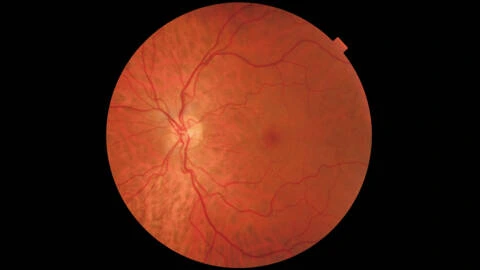
Captures detailed, accurate fundus images for reliable retinal assessments.
Fully Automated Operation
Robotic system enables automatic alignment, focus, and image capture for consistent results.
Small Pupil Adaptability
Acquires clear images from pupils as small as 3.3mm, improving accessibility for more patients.
Brochures & Documentation
High-Quality Imaging with Simple Operation
Fully Automated Mode
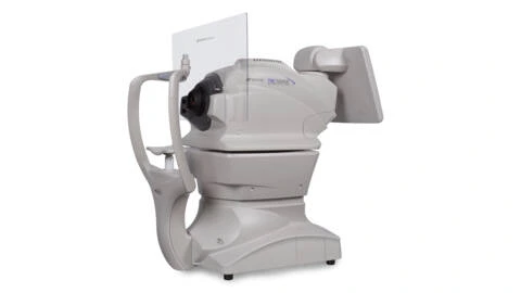
The TRC-NW400 offers a fully automatic mode where alignment, focusing, image capture, and switching between eyes are performed automatically. With minimal training required, the operator simply selects the mode, centers the pupil on the screen, and presses “Capture START” to obtain high-quality images consistently.
Manual Operation Mode
For greater control, manual mode allows the operator to adjust the camera position, illumination, flash intensity, diopter settings, and fixation targets using the touchscreen interface. This is particularly useful when focusing on specific areas of the fundus.
Compact and Space-Saving Design
The rotating touch panel monitor enables flexible operation from any angle, allowing the practitioner to position the device in front, behind, or beside the patient. Its compact footprint makes it suitable for clinics with limited space and allows operation even when placed against a wall or in a corner.
Low Flash Intensity for Patient Comfort
A low-intensity 4.0Ws flash minimizes patient discomfort while maintaining high-quality image capture. This reduces strain and enables multiple images to be taken efficiently.
Small Pupil Imaging and Zoom Function
The TRC-NW400 can capture retinal images from pupils as small as 3.3mm. Features such as small pupil mode, digital zoom, and an overlay mask help optimize image acquisition, automatically adjusting settings for improved imaging in patients with smaller pupils.*
*Settings may vary depending on configuration.